


Seeing Synapses
Synapses, the links between neurons that allow them to pass information to one another, are central to all the brain’s functions. These key building blocks are believed to be where memories are stored, yet researchers have little understanding of how they change when memories are made. New insights into the role of synapses in memory, learning and forgetting — or how sleep, drugs and other stimuli affect them — would revolutionize scientific understanding of the entire brain.
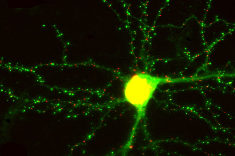
At present, researchers cannot image the entire brain at a high enough resolution to see synapses, tiny structures only a hundredth the diameter of a human hair. That’s partly because scientists have yet to build the technology needed to selectively label, image and analyze these key but miniscule structures.
The multidisciplinary team of distinguished USC scientists Scott Fraser, Don Arnold and Carl Kesselman aim to change that.
 The trio has received a highly competitive five-year, $9.7-million Transformative Research Grant from the National Institutes of Health to perform the first direct study of living synapses in the intact brain, allowing researchers to analyze synaptic changes in zebrafish as a memory is made. The USC researchers chose zebrafish because their transparent brains make them easier to image. They are also large enough to show the same complexities, mechanisms and diseases as the human brain, including mental illness, making the results relevant to neuroscientists and other health researchers.
“We want to create a new way of observing how the brain functions in a way that nobody has ever done before,” Kesselman said. “Our approach will offer an overall map of the brain’s synaptic changes, creating an entire circuitry diagram compared to just a view of a single wire.”
Such a comprehensive picture of the brain’s synapses could one day lead to a better understanding of and treatments for post-traumatic stress disorder (PTSD) and other neurological conditions in humans, he added.
The zebrafish experiments will use a new class of genetically embedded probes developed by Arnold of the USC Dornsife College of Letters, Arts and Sciences.
Fraser,a provost’s professor with appointments from the USC Viterbi School of Engineering, USC Dornsife, and the Keck School of Medicine of USC, is working with an interdisciplinary team to build a powerful light sheet microscope optimized to image the synapses that Arnold’s team will label. The microscope is so fast and sensitive that it can capture a comprehensive image of a zebrafish’s synapses before, during and after a memory is made.
“We pride ourselves on building microscopes that redefine the impossible,” said Fraser, one of the world’s foremost imaging experts.
Kesselman — a dean’s professor who also serves as director of the Informatics Division at the USC Information Sciences Institute (ISI) — has designed algorithms that will identify, sort and store zebrafish synapses, a challenging proposition given their small size, large number and amount of data. These huge databases of information “will be accessible to researchers using the Big Data tools the USC researchers are developing,” Kesselman said.
Added Arnold: “People have wanted to see this [synaptic changes] for the last 100 years, but the technology wasn’t there — until now.”