


MRI Machine – One of Just Three in the World – Arrives on Campus
WHEN MAGNETIC-RESONANCE imaging machines first came into clinical use in the 1980s, they represented one of the biggest advancements in medical imaging technology. But as valuable as these machines are, they are not without their shortcomings. Traditional MRI machines struggle to produce reliable images of certain body parts, such as the heart, lungs, and vocal tract, and of body parts that are near metallic implants. They are also exceedingly large and expensive.
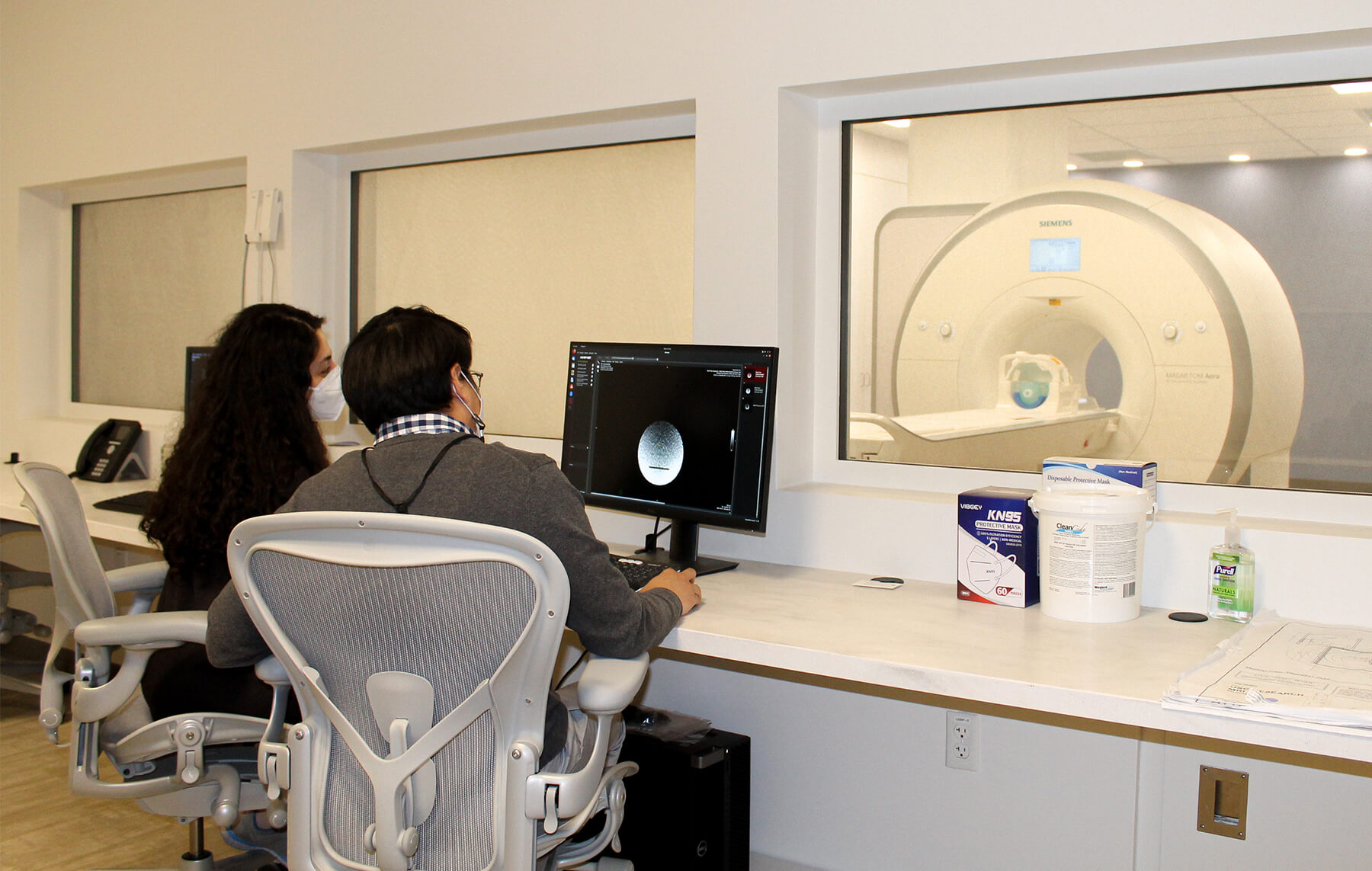
Thanks to an initiative led by Krishna Nayak, USC Viterbi professor of electrical and computer engineering, a new type of MRI machine was installed in December 2019 in the USC Michelson Center for Convergent Bioscience. In fact, this machine is one of only three of its kind in the world. Known as a high-performance, low-field MRI, it produces better images of certain body parts while more reliably handling patients with implants, in a much quieter environment. It also has the potential to bring MRI technology to communities and facilities that cannot afford traditional MRI machines.
“Low-field MRI has the potential to drive down the system cost and physical footprint so that MRI technology can be more accessible and reach more patients in need,” said Nayak. “There will be many engineering challenges, and we look forward to solving them.”
The machine will be used collaboratively by a cross section of researchers from USC Viterbi, USC Dornsife, the Keck School of Medicine of USC and Children’s Hospital Los Angeles.
One of those collaborators is Dr. John Wood. A professor of pediatrics and radiology at Keck USC, Wood also serves at CHLA and has a background in electrical and biomedical engineering. For him, the ability to better conduct real-time imaging cannot be understated. “A normal MRI can take up to 13 seconds to profile one slice of the heart. When you’re trying to understand how well a child’s heart is beating, that’s simply too slow,” he said.
High-performance, low-field imaging could have a big impact for young patients who have pacemakers or vascular hardware that interfere with traditional MRI scans, Wood said. He is now working on a grant to use the new machine for improved fetal imaging.
Dr. Jay Acharya, a professor of radiology at USC Keck, where he serves as director of Spine Imaging Intervention, says he cannot overstate the value of working with USC Viterbi engineers like Nayak.
“Without the input and skillset of people like Krishna, there’s a lot I wouldn’t be able to do for my patients. The harmonization of the two schools within the USC system is so important. Neither one of us could do this on our own,” Acharya said.
“Although Krishna is an engineer, he possesses a great deal of medical insight, and I’ve appreciated the cross-fertilization of our work over the years,” Wood added.
Low-field MRI might have an even larger economic impact on society in the future. The weaker magnet used in low-field MRI means these machines could cost much less and become much smaller and easier to maintain.
Funded by generous support from the USC Viterbi School of Engineering and a National Science Foundation grant, the machine’s home in Michelson Hall is appropriate, as it represents a true convergence of engineering and medicine. In addition to lead PI Nayak, the project has four co-PIs: fellow USC Viterbi faculty Shri Narayanan and Justin Haldar, and Dani Byrd and Khalil Iskarous, both professors of linguistics at USC Dornsife.
For Narayanan, a 20-year veteran of USC Viterbi’s Signal and Image Processing Institute, the project is a natural progression of his department’s leadership in the field. “The installation of the low-field MRI scanner ushers in a new and an immensely exciting phase in our two-decades-long program on developing and using real-time imaging to the scientific and clinical study of human function,” he said.