


This is Your Child’s Brain on Bach… in 3-D
USC researchers — in a partnership with the Los Angeles Philharmonic Association, the nonprofit Heart of Los Angeles and Youth Orchestra LA — have found that auditory pathways in the brains of young children receiving classical music training are maturing more quickly than those of other children in the study. Additionally, the young musicians are more accurate at detecting pitch changes in the melodies.
“The auditory system is stimulated by music,” said Assal Habibi, lead author of the recent results published in the journal Developmental Cognitive Neuroscience, and a senior research associate at the Brain and Creativity Institute at USC Dornsife College of Letters, Arts and Sciences. “This system is also engaged in general sound processing that is fundamental to language development, reading skills and successful communication.”
Explained Justin Haldar, USC Viterbi assistant professor in the Ming Hsieh Department of Electrical Engineering: “In some ways the brain is like a muscle. If you exercise it, then the regions that you’re exercising will get stronger. And the process of getting stronger can be associated with detectable changes to the brain tissue at the microscopic level.”
Anecdotal evidence has long suggested a correlation between musicianship and creativity, as well as musicianship and scientific ability. Past research on the impact of music on the brain has largely focused on adults. However, the USC–LA Philharmonic–HOLA developmental study follows 65 children ages 6 or 7 for five years, comparing brain changes among three groups: 21 children who receive ongoing classical music instruction through HOLA’s youth orchestra; 24 in a community soccer program; and 20 not involved in any specific after-school activities.
The initial reported findings were based on a technique called EEG that measures the electrical activity of the brain. Now, specialized software created by a USC Viterbi professor and his former Ph.D. student, both from the Ming Hsieh Department of Electrical Engineering, is allowing the researchers to analyze MRI data to see differences in children’s brain structure as they emerge.
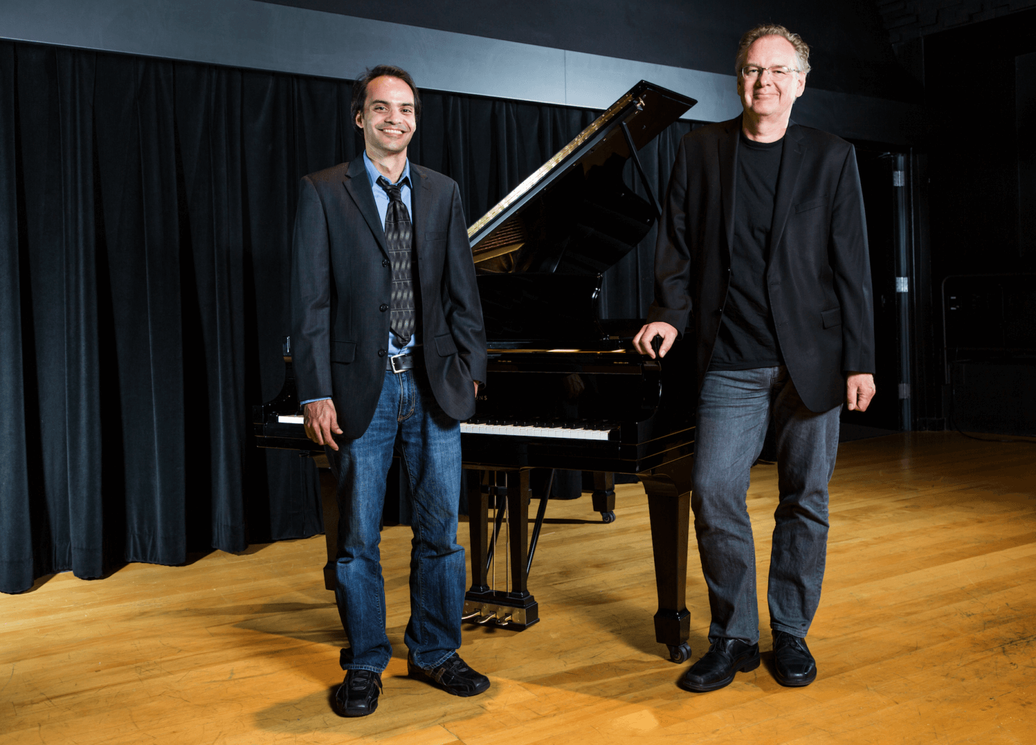
BrainSuite, software co-created by Richard Leahy, a USC Viterbi professor of electrical engineering, and David Shattuck, now a professor at UCLA, has many advantages over competing products, said Hanna Damasio, director of the Dornsife Cognitive Neuroscience Imaging Center and a project researcher.
“BrainSuite has been our choice of tool to investigate structural changes because it is an automated engine,” Damasio said. “It allows us to study the development of cortical thickness in specific brain regions, as well as the anatomical connectivity between key cortical areas. Additionally, BrainSuite’s results can be inspected at every step, allowing us to [modify], whenever necessary, to optimize the analysis.”
Haldar added that analyzing the MRI data with BrainSuite “will provide a more complete picture of the trajectory of brain development.”
BrainSuite’s capabilities are not limited to music research. The software helps researchers in a number of disciplines, from neuroscience to radiology, by providing them with a tool through which they can explore the brain and its role in human development, cognition, behavior and disease.
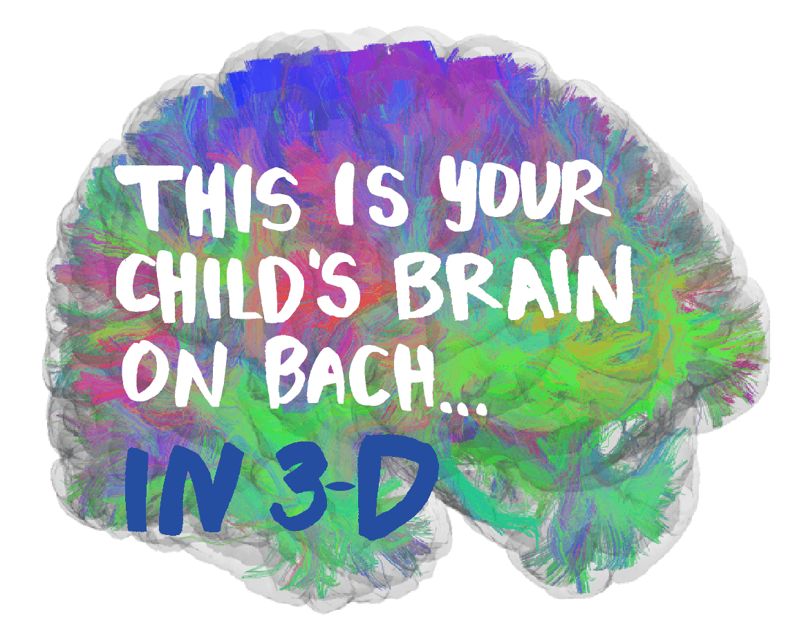
Its 3-D images depict a colorful, exquisite landscape that an ordinary onlooker might mistake for art. But to a scientist, the images map the condition and composition of the human brain.
BrainSuite takes 3-D images of the brain and can morph each of these images onto a common representation of the brain, or an atlas.
BrainSuite enables researchers to track changes in brain size, shape or function. The open-source software also allows researchers to compare the brain images of different study subjects, and to compare specific areas in one subject’s brain to another’s.
“We can analyze, for example, whether there are regional differences in the size or shape of the brains of patients with Alzheimer’s disease and those with mild cognitive impairment,” Leahy said. “More generally, we can detect differences between a control group and different disease populations. In a similar manner, BrainSuite can also be used to investigate differences between groups addressing, for example, questions of nature versus nurture, and the impact of genetics and the environment on the brain.
“BrainSuite is able to visualize and analyze the brain’s white matter,” Leahy added, “which contains the axonal fibers or ‘wiring’ between different brain areas, to better understand how the brain functions as a complex set of interacting networks.”
In addition to analyzing conventional MRI scans, BrainSuite can perform automated diffusion MRI analysis, which maps the movement of water molecules in the brain. Diffusion MRI allows scientists to study the microscopic features of brain tissue that are too small to see with conventional MRI. Such microscopic changes may be associated with aging and disease, or the subtle changes that manifest after learning something new. BrainSuite is a key tool for current scientific studies at USC.
“We are using BrainSuite for an NIH-funded study on chronic stroke survivors,” said Carolee Winstein, a professor at USC’s Division of Biokinesiology and Physical Therapy. “The software helps us to measure the integrity of the corticospinal tract, which controls the movement of skeletal muscles. We hope to determine if changes in the corticospinal tract beyond six months of a stroke are associated with improvements in movement for patients who have suffered mild to moderate impairment.”
In another study, researchers at the Keck School of Medicine of USC are working with Rancho Los Amigos National Rehabilitation Center and the Cleveland Clinic to study a type of epilepsy caused by a birth defect, focal cortical dysplasia, where neurons in an area of the brain have failed to migrate to the proper formation of the brain. FCD disrupts brain circuitry and causes seizures.
Many cases of FCD are difficult to detect through imaging. Saman Hazany, an assistant professor of neuroradiology at the Keck School of Medicine, said scientists are working with Leahy to test BrainSuite and see if it improves FCD detection so that doctors can treat those patients appropriately.
And then there’s the music study.
“Intensive music training during childhood may have a broader and lasting impact, particularly in those from disadvantaged backgrounds,” Leahy said. “This study will help us to better understand the effects of early-music training, possibly leading to new insights into the impact and importance of music on brain development.”
Managing editor Marc Ballon contributed to this story.